(a) Rectangular stitching leads to seams (black lines) going through the many microlenses, whereas adaptive stitching puts the seams at the boundaries of the microlenses to mitigate artefacts. They validated the reconstructed outcomes in comparison with two-photon microscopy to understand limits of the new technique.

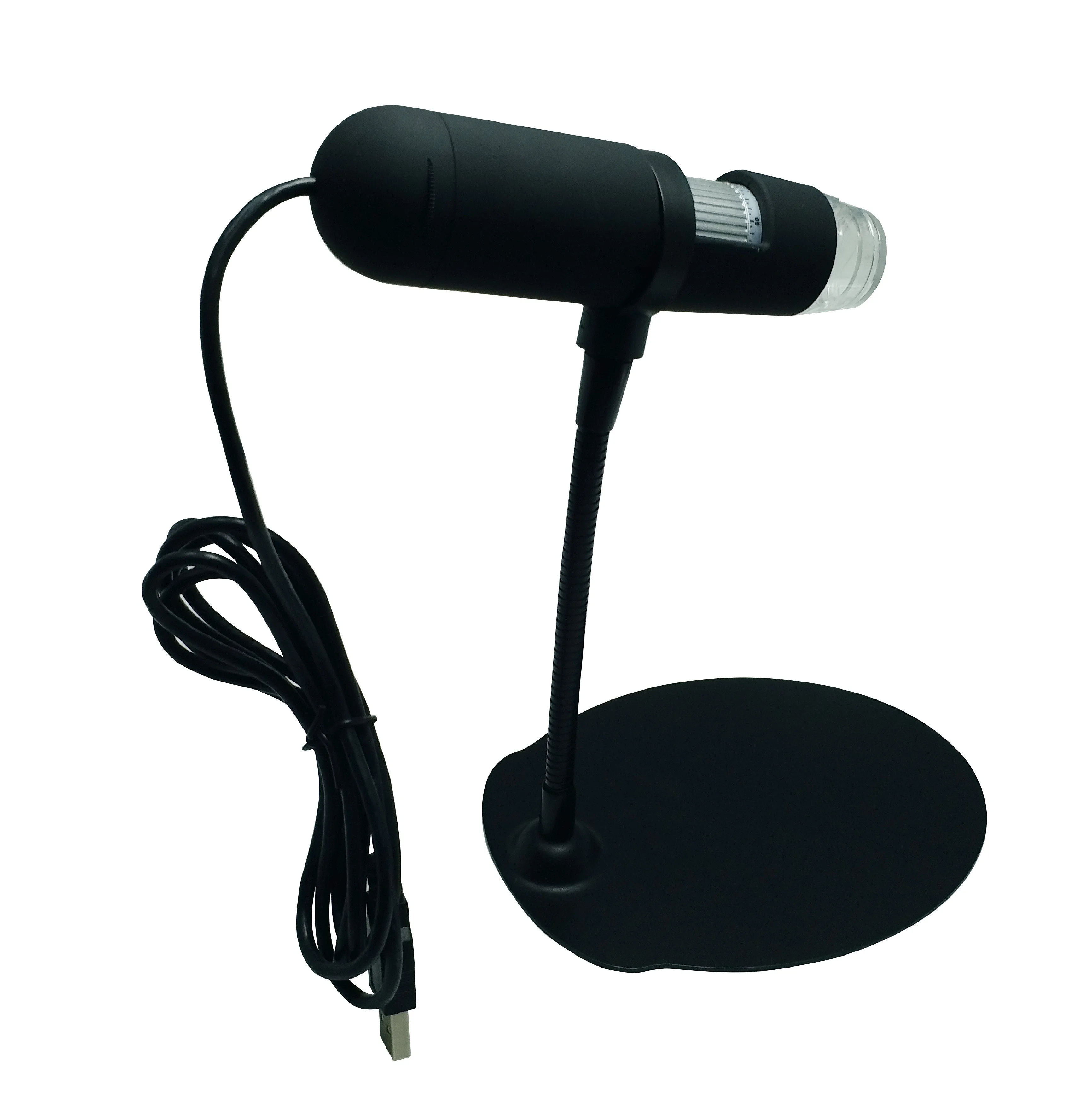
The team tested the microscopic capabilities by imaging fluorescent resolution targets as well as freely swimming biological samples and mouse brain tissue. developed a 3-D miniscope to achieve higher resolution with lighter weight compared to existing techniques. For example, a previously developed miniature light-field microscope (MiniLFM) can process neural activity with an optimized algorithm. Single-shot methods can enable faster capture speeds and a temporal resolution limited by the camera frame rate. Such microscopes are also known as "miniscopes" and are made of 3-D printed parts, although offering 2-D fluorescence imaging alone. Miniature fluorescence microscopes are important in systems biology for optical recordings of neural activity in free-moving animals, long-term in situ imaging in incubators and medical devices. Miniature fluorescence imaging and technical innovations optically recorded neural activity in free-moving animals and in long-term in situ imaging applications in incubators and within lab-on-a-chip devices. Using the device, Yanny and Antipa et al. They engineered the new device known as the Miniscope3D by replacing the tube lens of a conventional 2-D miniscope with an optimized multifocal phase mask at the objective's aperture stop. In a new report now on Nature Light: Science & Applications, Kyrollos Yanny, Nick Antipa and a team of scientists in the Joint Graduate Program in Bioengineering, Electrical Engineering and Computer Sciences at the University of California, Berkeley and the Universite libre de Bruxelles Belgium, developed a single-shot 3-D fluorescence microscope. Existing miniature fluorescence microscopes are a standard technique in life sciences, but they only offer two-dimensional (2-D) information. Credit: Light: Science & Applications, doi: 10.1038/s41373-7Ī miniature fluorescence microscope that weighs less while offering high resolution compared to existing devices will have a range of applications in systems biology. The 3D reconstruction here is of a freely swimming fluorescently tagged tardigrade. The forward model is then used to iteratively solve an inverse problem to reconstruct 3D volumes from single-shot 2D measurements. We use this data set to pre-compute an efficient forward model that accurately captures field-varying aberrations. A sparse set (64 per depth) of calibration point spread functions (PSFs) is captured by scanning a 2.5 μm green fluorescent bead throughout the volume. We remove the Miniscope’s tube lens and place a 55 μm thick optimized phase mask at the aperture stop (Fourier plane) of the GRIN objective lens.


Compared with previous Miniscope and MiniLFM designs, our Miniscope3D is lighter weight and more compact.


 0 kommentar(er)
